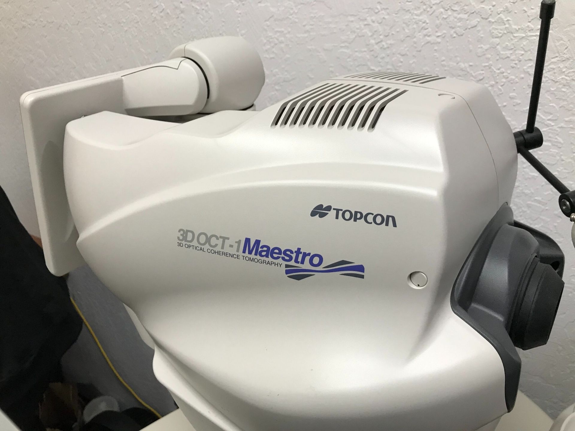
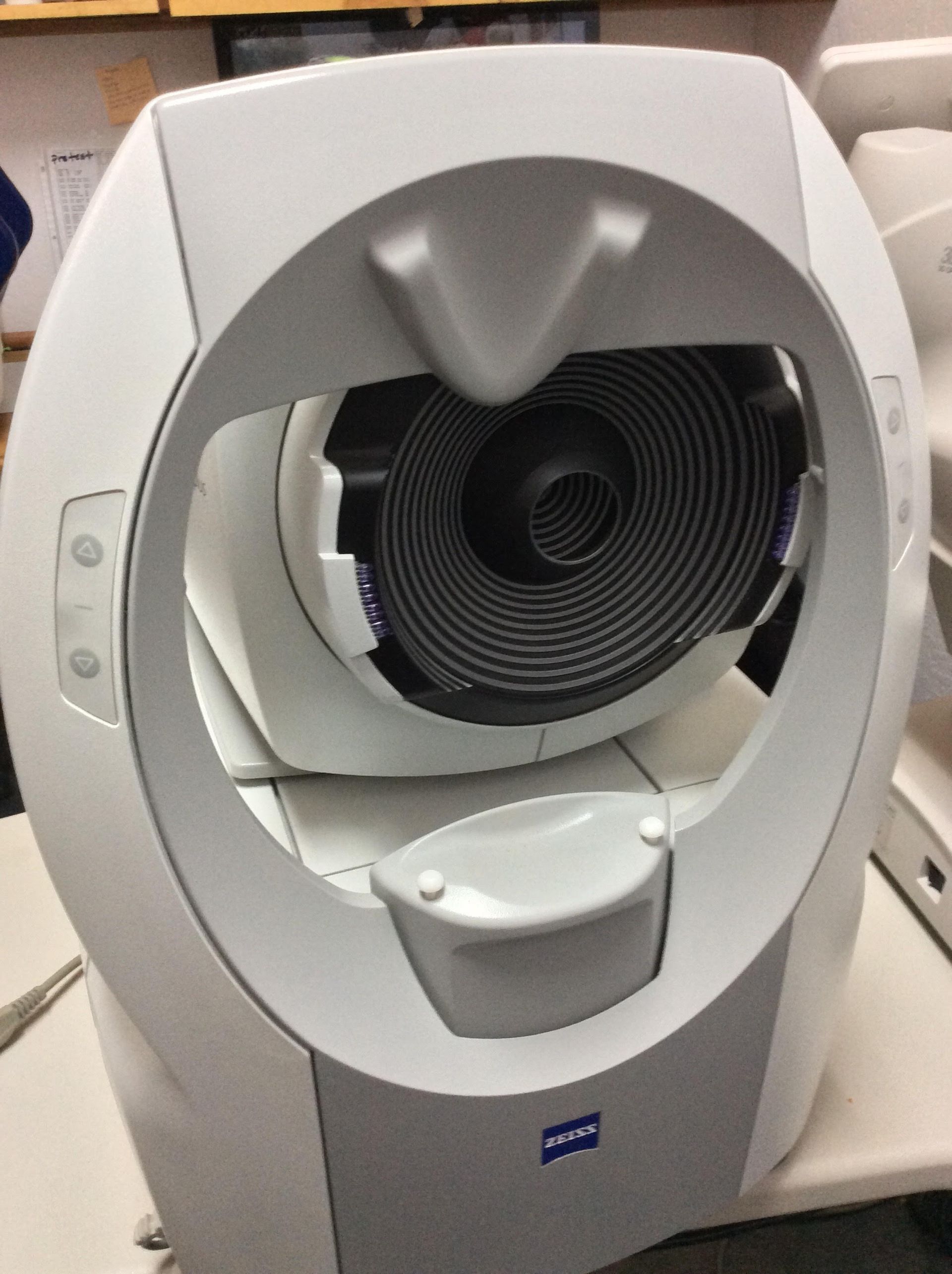
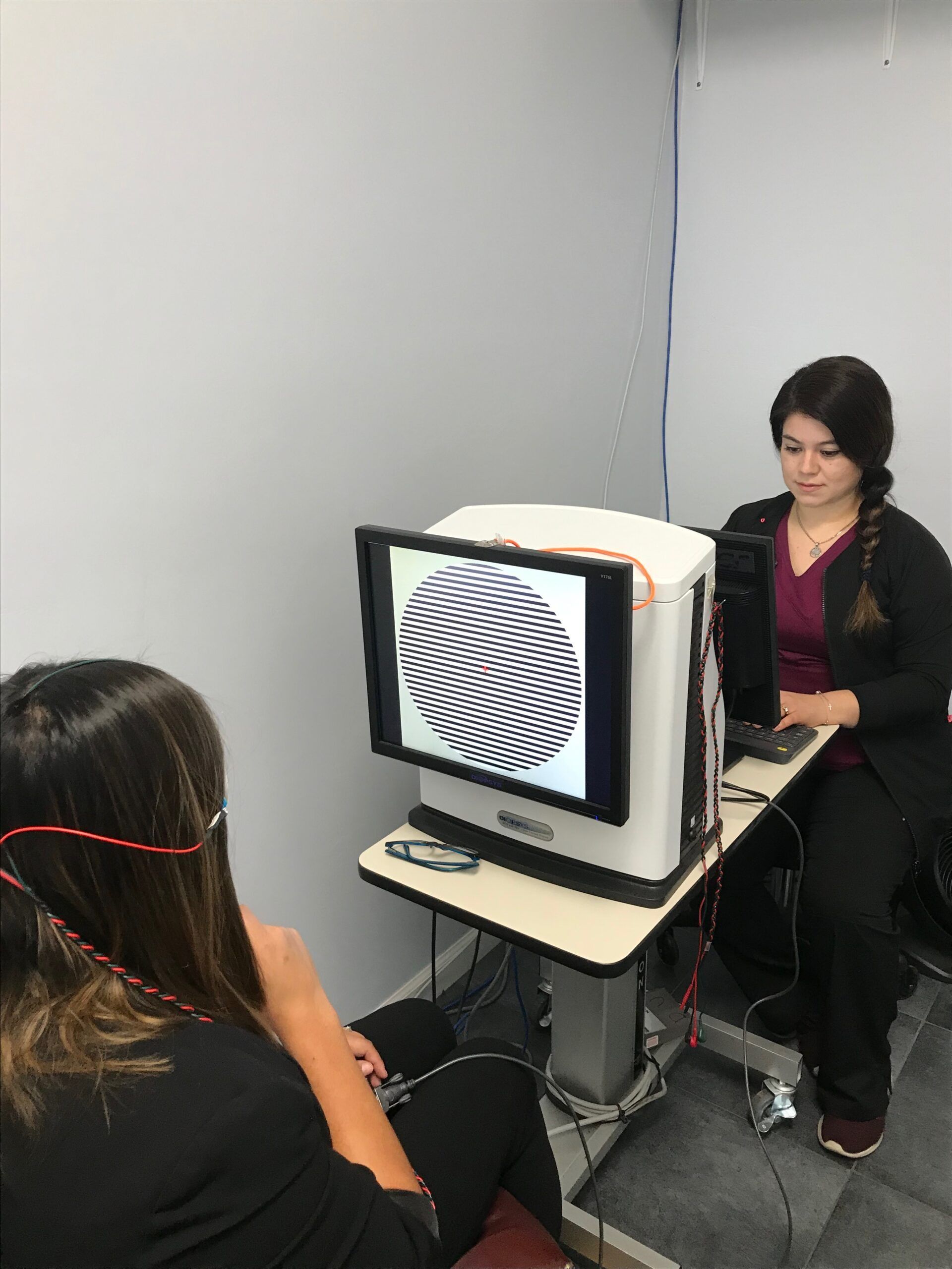
If your doctor recommended OCT or retinal imaging, it is because parts of your eye cannot be evaluated well from the outside.
Both are quick, neither touches your eye, and together they let us examine the back of the eye and document what we find.
What retinal imaging shows
A retinal photograph is a wide, documented view of the back of your eye: the retina, the optic nerve, and the blood vessels running across them.
Its real value is that it is a record. A photo from today can sit beside one from three years ago, and gradual change that no one could hold in memory becomes visible.
What OCT adds
A photograph shows the surface. OCT shows depth.
OCT produces cross-sectional images of the retina, so we can examine its individual layers rather than only the view from above. It helps us look for swelling, thinning, and structural changes that a photograph alone would not reveal.
Think of the photograph as looking down at the ground, and OCT as looking at the layers in a core sample.
When we recommend them
Imaging is commonly useful in the context of:
- Diabetes, where retinal blood vessels can be affected before vision changes
- High blood pressure, which also shows up in retinal vessels and covered in how high blood pressure affects your eyes
- Macular conditions, where central retinal layers matter
- Glaucoma monitoring, where clinically appropriate, to examine the optic nerve and nerve fiber layer
It is also useful simply to establish a baseline, so future comparisons have a starting point.
Not every patient needs imaging at every visit. The doctor recommends it based on your symptoms, exam findings, and health history.
What the appointment is like
- You rest your chin on a support and look at a target inside the instrument.
- The scan runs. It takes seconds per eye.
- Nothing touches your eye and nothing is uncomfortable. You may see a bright light or a moving line.
- The doctor reviews the images with you and explains what he is looking at.
Dilation is sometimes recommended for a clearer look at the retina, though many scans can be done without it. A dilated exam is also how we check the retina after new flashes and floaters.
What imaging does not do
Imaging supports the doctor's evaluation. It does not diagnose a condition on its own, and it does not replace his judgment.
It gives him more detail to work with, and it documents your eyes in a way that makes next year's exam more informative.
Common questions
Is there radiation?
No. OCT uses light, not radiation.
Does it hurt?
No. Nothing touches your eye.
Will insurance cover it?
It depends on the reason for the test and your plan. Imaging ordered for a medical reason is often billed to medical insurance. Our team can help you understand what applies.
Can I see the images?
Yes, and we encourage it. Most people find their own retina more interesting than they expected.
How often would I need this?
It depends on what we are monitoring. For some conditions annually, for others only once to establish a baseline.
Schedule an exam
Request an appointment or call 805-925-9575.